3d imaging-soft-tissue samples: x-ray specific staining method l protocol preview
Published 2 years ago • 17 plays • Length 2:01Download video MP4
Download video MP3
Similar videos
-
 2:01
2:01
high resolution 3d imaging: biological samples by micro ct l protocol preview
-
 2:01
2:01
3d u/s imaging: morphometry of muskoskeletal tissue | protocol preview
-
 2:01
2:01
visualization of 3d white adipose tissue structure l protocol preview
-
 2:01
2:01
adipose tissue structure by methylsalicylate clearing & 3d imaging l protocol preview
-
 2:01
2:01
horizontal whole mount: processing and imaging of 3d tissue of skin | protocol preview
-
 2:01
2:01
3d printing of preclinical x-ray computed tomographic data sets l protocol preview
-
 2:01
2:01
microct imaging of cnidaria, annelida, and xenacoelomorpha | protocol preview
-
 2:01
2:01
confocal time lapse imaging: cytocompatibility of dental composites | protocol preview
-
 9:57
9:57
cbct positioning
-
 10:46
10:46
microscope tutorial - how to stain with h&e
-
 7:41
7:41
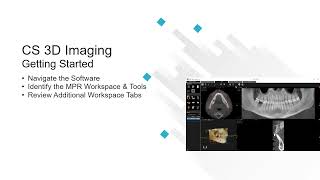
cs 3d imaging: getting started recorded class
-
 2:01
2:01
high resolution 3d imaging of human pancreas neuro-insular network l protocol preview
-
 2:01
2:01
tissue engineering-human 3d tumor test system l protocol preview
-
 2:01
2:01
elastic staining on slides of gastric cancer tissue | protocol preview
-
 2:01
2:01
serial block-face scanning electron microscopy: biological tissue samples l protocol preview
-
 2:01
2:01
visualizing proteins & macromolecular complexes: negative stain em l protocol preview
-
 2:01
2:01
extracting metrics for 3d root systems: x-ray ct data analysis | protocol preview
-
 2:01
2:01
biomedical tomographic imaging data & associated labels-ssle: glass crystals l protocol preview
-
 2:01
2:01
biplanar videoradiography: measuring 3d in-vivo shoulder kinematics | protocol preview
-
 2:01
2:01
light-sheet microscopy for 3d visualization of immune cells | protocol preview
-
 2:01
2:01
ct angiography for aortic endoleaks and 2d-3d imaging treatment | protocol preview
-
 2:01
2:01
3d human skin reconstruct model: tool to study normal skin & melanoma progression l protocol preview