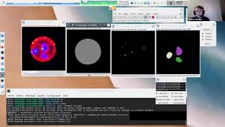
cell volume and morphology analysis with 3dsuite (imagej)
Published 2 years ago • 3.8K plays • Length 9:15Download video MP4
Download video MP3
Similar videos
-
 10:45
10:45
fiji (imagej): morphology & network analysis of mitochondria
-
 1:41
1:41
how to analyze cell culture spheroids: a step-by-step guide
-
 7:48
7:48
i2k 2020 tutorial: 3d analysis with the 3d imagej suite (session 1, part 1)
-
 5:16
5:16
fiji (imagej): using 3d objects counter and 3d manager to measure cells in a z stack
-
 41:36
41:36
a beginners guide to imagej (and fiji)
-
 24:41
24:41
morphology analysis using image j
-
 18:57
18:57
morphological segmentation with fiji (imagej)
-
 1:59
1:59
olympus novisight™ | 3d cell analysis system
-
 7:46
7:46
i2k 2020 tutorial: 3d analysis with the 3d imagej suite (session 1, part 3)
-
 1:15:11
i2k 2020 tutorial: 3d analysis with the 3d imagej suite (session 1, part 2)
-
 3:46
3:46
how to count number of cells, measure volume surface area & mean intensity of 3d stack image in fiji
-
 0:22
0:22
imagej automated spheroid analysis macro at work
-
 2:26
2:26
incellis cell imager – overview
-
 3:20
3:20
how to count the cells in tissue sections using imagej
-
![introduction to 3d analysis with 3d imagej suite [neubias academy@home] webinar](https://i.ytimg.com/vi/OPC2kP-5By4/mqdefault.jpg) 1:47:33
1:47:33
introduction to 3d analysis with 3d imagej suite [neubias academy@home] webinar
-
 9:38
9:38
3d mitochondria analysis using imagej: (part1)
-
 1:06:38
i2k 2020 tutorial: 3d analysis with the 3d imagej suite (session 2, part 1)
-
 44:07
44:07
i2k 2020 tutorial: 3d analysis with the 3d imagej suite (session 2, part 3)
-
 57:06
57:06
i2k 2020 tutorial: 3d analysis with the 3d imagej suite (session 2, part 2)