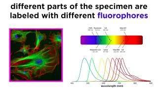
confocal imaging of cell cytoskeletal structures
Published 11 years ago • 526 plays • Length 0:13Download video MP4
Download video MP3
Similar videos
-
 3:31
3:31
confocal microscopy
-
 27:16
27:16
introduction to confocal microscopy
-
 17:42
17:42
david albrecht: nanoscale imaging of live cells with confocal interferometric scattering microscopy
-
 0:38
0:38
ixplore spin | confocal imaging of rapid cell dynamics
-
 9:43
9:43
widefield and confocal fluorescence microscopy
-
 18:14
18:14
confocal microscopy | what is the difference between confocal and fluorescence microscopy?
-
![what is a confocal microscope - webinar [leica microsystems]](https://i.ytimg.com/vi/XAiNE5iYMas/mqdefault.jpg) 35:32
35:32
what is a confocal microscope - webinar [leica microsystems]
-
 8:23
8:23
confocal microscope
-
 0:13
0:13
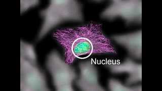
live-cell confocal imaging showing the continuous change of the actin cap
-
 3:31
3:31
opterra high-speed live cell imaging multipoint scanning confocal microscope
-
 13:57
13:57
multiple imaging modes for enhanced analysis of drosophila cells with the leica sp8 confocal system
-
 1:14:03
uncovering the membrane mechanism of cytokinesis using live cell imaging and subcellular...
-
 0:23
0:23
real time confocal imaging of lh-induced endocytosis of flag-tagged lh receptor in hek 293 cells.
-
 9:17
9:17
cytoskeleton structure and function
-
 0:59
0:59
confocal microscopy in 1 minute | principles of confocal microscopy | application of confocal
-
 0:05
0:05
f-actin live cell staining with sir-actin in cardiac myocytes
-
 24:47
24:47
microscopy: optical sectioning and confocal microscopy (kurt thorn)
-
 0:02
0:02
comparison of confocal and sted time-lapse movies of vimentin and tubulin dynamics
-
 0:24
0:24
the confocal microscope: a brief introduction pt 1 🔬 #shorts | terasaki institute
-
 44:11
44:11
introduction to confocal microscopy
-
 12:18
12:18
confocal microscopy principle tutorial
-
 0:03
0:03
bioflux system: confocal image of a biofilm showing 3d structure