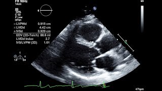
echocardiographic anatomy part 2: psax (short axis)
Published 2 years ago • 4.8K plays • Length 1:56Download video MP4
Download video MP3
Similar videos
-
 11:21
11:21
echocardiographic anatomy; part 1: plax (parasternal long axis)
-
 0:47
0:47
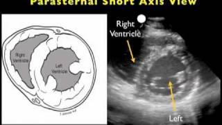
parasternal short axis (psax)
-
 1:12:51
transthoracic echo full protocol. part ii: parasternal view (plax , psax, rvit, rvot, m-mode)
-
 3:47
3:47
case study 2: echocardiographic anatomy
-
 1:48
1:48
echocardiographic anatomy part 4: apical two chamber(a2c)
-
 4:30
4:30
routine echocardiogram protocol with standard 2d echo images and color doppler
-
 25:44
25:44
how to perform standard echo measurements
-
 16:18
16:18
cardiac structures! (apical 5, 2 and 3 chamber views - echocardiography)
-
 9:22
9:22
anatomy of heart in echo part 1: location & orientation
-
 1:00
1:00
2 d- measurements in echo- psax
-
 2:02
2:02
parasternal short axis view - transthoracic echocardiography (ultrasound)
-
 10:13
10:13
echocardiographic anatomy part 6: right ventricle focused view (rv focused)
-
 0:46
0:46
right ventricle outflow tract (rvot)
-
 3:46
3:46
cardiac ultrasound - parasternal short axis - sonosite, inc.
-
 3:02
3:02
tips & mistakes: plax (parasternal long axis)
-
 5:38
5:38
echocardiography essentials: mastering the parasternal short axis (psax) view of the aortic valve
-
 4:48
4:48
wall motion abnormality part ii: cases & practice
-
 12:45
12:45
cardiac structures! - echocardiography (parasternal short axis view)
-
 5:03
5:03
hands-on 4: psax tips & tricks