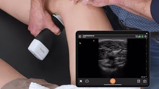
femoral vein doppler ultrasound normal vs abnormal image appearances | deep vein thrombosis usg scan
Published 1 year ago • 17K plays • Length 3:26Download video MP4
Download video MP3
Similar videos
-
 4:08
4:08
popliteal vein doppler ultrasound normal vs abnormal image appearances | deep vein thrombosis usg
-
 7:14
7:14
femoral vein doppler ultrasound probe positioning | lower limb veins usg scanning technique
-
 8:40
8:40
saphenous vein doppler ultrasound normal vs abnormal | varicose veins | lower limb vascular usg
-
 8:27
8:27
femoral artery doppler ultrasound normal vs abnormal | stenosis/occlusion/pseudoaneurysm/avf usg
-
 0:34
0:34
acute and chronic deep vein thrombosis. vein diameter variation | ultrasound
-
 0:20
0:20
normal & abnormal common femoral vein doppler
-
 1:27:14
abdominal ultrasound normal vs abnormal images | liver, gallbladder, pancreas, kidney, hernia usg
-
 14:00
14:00
peripheral artery ultrasound interpretation | 15 minute radiology cme
-
 38:38
38:38
varicose vein doppler demo: part 1
-
 1:24
1:24
deep vein thrombosis (dvt) - ultrasound scanning technique
-
 9:56
9:56
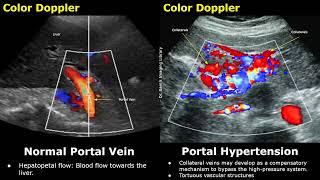
portal vein color & spectral doppler ultrasound normal vs abnormal images | liver vascular usg scan
-
 6:52
6:52
ultrasound tutorial: dvt / lower limb veins | radiology nation
-
 10:10
10:10
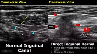
hernia ultrasound normal vs abnormal images | direct/indirect inguinal/epigastric/femoral hernia usg
-
 9:19
9:19
popliteal artery spectral/color doppler ultrasound normal vs abnormal images | vascular usg
-
 11:06
11:06
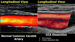
carotid artery color/spectral doppler ultrasound normal vs abnormal images | ica stenosis usg
-
 0:31
0:31
b-mode appearance of deep veins after deep vein thrombosis
-
 7:38
7:38
dvt or deep vein thrombosis affecting femoral and popliteal veins, ultrasound color doppler video
-
 7:01
7:01
doppler ultrasound imaging for detection of deep vein thrombosis in plastic surgery outpatients
-
 0:43
0:43
common femoral vein thrombosis colour doppler ultrasound video
-
 6:59
6:59
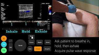
lower extremity dvt ultrasound examination