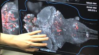
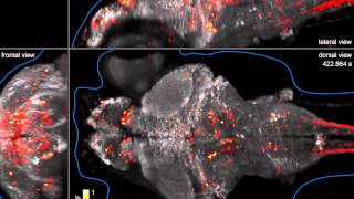
high resolution imaging of zebrafish at nano and millimeter scale using andor dragonfly
Published 3 years ago • 554 plays • Length 1:01:32Download video MP4
Download video MP3
Similar videos
-
 1:01:32
high resolution imaging of zebrafish at nano and millimeter scale
-
 0:51
0:51
in vivo imaging of blood flow in mouse intestine - andor dragonfly confocal
-
 0:16
0:16
live imaging of zebrafish vascular development with andor's bc43 benchtop microscope system
-
 0:41
0:41
new dragonfly 600 confocal microscope system
-
 0:16
0:16
beautiful imaging of zebrafish development
-
 7:12
7:12
confocal microscopy live imaging of zebrafish embryonic brain
-
 4:49
4:49
flashes of insight: whole-brain imaging of neural activity in the zebrafish
-
 9:44
9:44
looking for answers in the skull of a zebrafish
-
 2:01
2:01
using optokinetic response to study visual function of zebrafish l protocol preview
-
 1:19
1:19
how to: view zebrafish without injections
-
 0:20
0:20
zeiss lsm 880 with airyscan: fast mode calcium imaging of live zebrafish embryo
-
 1:40
1:40
mouse hippocampus expanded sample captured with andor dragonfly confocal and rendered in imaris
-
 0:34
0:34
whole-brain imaging of neuronal activity in a larval zebrafish
-
 4:14
4:14
dragonfly: the development story
-
 0:53
0:53
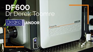
dragonfly confocal - customer testimonial - dr derek toomre - yale school of medicine
-
 0:17
0:17
scape 2.0 dual-color 3d imaging of zebrafish embryo and c. elegans.
-
 12:22
12:22
two-photon axotomy and time-lapse confocal imaging in live zebrafish embryos
-
 0:11
0:11
optimized multipoint confocal systems deliver optical sectioning up to mm inside a tick tissue.
-
 0:04
0:04
adhesion protein visualized by tirf microscopy using andor dragonfly confocal microscope
-
 0:26
0:26
imaging zebrafish at mit
-
 0:09
0:09
seamless zebrafish intestine stitched microscopy image acquired with bc43 benchtop microscope system