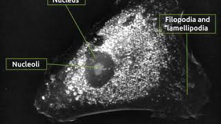
morphological evolution of cortical neuron cells over time under nanolive live cell imaging solution
Published 3 years ago • 1.1K plays • Length 3:00Download video MP4
Download video MP3
Similar videos
-
 0:16
0:16
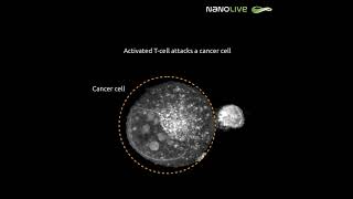
label-free live cell imaging: activated t-cell killing cancer cell
-
 0:16
0:16
activated t cell attacks a cancer cell
-
 0:42
0:42
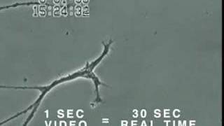
primary cortical neuron imaged with nanolive 9 days after addition of neurite stimulation media
-
 0:26
0:26
unlocking the mysteries of neurite growth in primary cortical neurons: a quantitative approach
-
 0:28
0:28
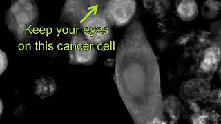
label-free live cell imaging: t-cells killing cancer cells - zoomed-in
-
 5:01
5:01
how mercury causes brain neuron damage - uni. of calgary
-
 13:10
13:10
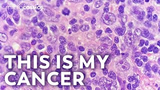
what does cancer look like?
-
 1:04
1:04
actual footage of cell division (kidney cells)
-
 0:31
0:31
nanolive imaging: failed mitosis and apoptosis by multinucleated mouse pre-adipocyte
-
 0:16
0:16
idorsia - differentiation of luhmes neuronal precursor cells into mature dopaminergic neurons
-
 0:29
0:29
is it possible to capture horizontal mitochondrial transfer in real time using nanolive imaging?
-
 0:36
0:36
stem cell differentiating into neuronal cells
-
 0:17
0:17
label-free live cell imaging of mesenchymal stem cells undergoing mitosis
-
 0:26
0:26
nanolive imaging: effects of excitation light (fitc 40%) - without fluorescent marker
-
 0:42
0:42
automated live cell imaging: the cx-a
-
 0:41
0:41
unstimulated hcn2 cells imaged under nanolive label-free live cell imaging
-
 0:17
0:17
human umbilical vein endothelial cells undergoing mitosis with nanolive imaging
-
 0:41
0:41
label-free live cell imaging of keratinocytes
-
 0:23
0:23
label-free live cell imaging of apoptosis of t685a human melanoma cancer cell
-
 0:15
0:15
label-free live cell imaging of simultaneous, massive mammalian cell necrosis
-
 0:26
0:26
nanolive imaging: effects of excitation light (fitc 20%) - without fluorescent marker
-
 0:13
0:13
nanolive imaging: effects of excitation light (dapi 10%) - without fluorescent marker