normal skin histology - explained by a dermatopathologist
Published 7 years ago • 289K plays • Length 1:14:19Download video MP4
Download video MP3
Similar videos
-
 16:23
16:23
skin histology: epidermis layers (stratum basale, spinosum, granulosum, lucidum & corneum)
-
 11:47
11:47
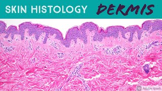
skin histology: dermis (the epidermis can't live without it!)
-
 59:59
59:59
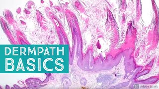
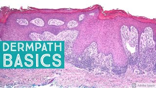
basic dermpath cases - explained by a dermatopathologist
-
 40:10
40:10
basic dermpath cases - explained by a dermatopathologist
-
 37:41
37:41
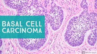
basal cell carcinoma (bcc) 101 - dermpath basics explained by a dermatopathologist
-
 47:01
47:01
basic dermpath cases - explained by a dermatopathologist
-
 47:40
47:40
basic dermpath cases - explained by a dermatopathologist
-
 40:56
40:56
basic dermpath cases - explained by a dermatopathologist
-
 32:32
32:32
my melanoma story | navigating stage 4 cancer as a mom of 2
-
 1:08:40
skin adnexal tumors: dermpath board review for dermatology pathology & dermpath
-
 13:40
13:40
nodular fasciitis - explained by a soft tissue pathologist
-
 21:29
21:29
sweat glands under microscope (skin adnexa histology anatomy eccrine apocrine sebaceous hair)
-
 9:46
9:46
skin histology: acral skin (aka glabrous skin of palm of hand & sole of foot)
-
 14:51
14:51
syringocystadenoma papilliferum (scap) 101...explained by a dermatopathologist
-
 18:35
18:35
cutaneous horn...explained by a dermatopathologist
-
 43:09
43:09
skin adnexal tumors 101: a basic approach for general pathologists
-
 36:09
36:09
dermatofibroma 101 (benign fibrous histiocytoma)...explained by a dermatopathologist
-
 4:25
4:25
melanoma vs nevus: microscopic clues for malignancy explained in 5 minutes
-
 20:13
20:13
primary cutaneous pecoma...explained by a dermatopathologist
-
 47:01
47:01
vasculitis...explained by a dermatopathologist
-
 9:19
9:19
hair follicle under microscope & scalp skin histology (dermatology dermpath pathology anatomy)
-
 20:16
20:16
immunohistochemistry in normal skin: p63, ema, desmin, sma, cd34, factor xiiia