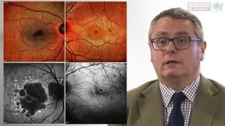
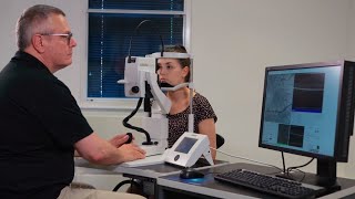
taking oct to the edge: evaluating the peripheral retina with oct
Published 1 year ago • 650 plays • Length 59:40Download video MP4
Download video MP3
Similar videos
-
 56:58
56:58
ordering and interpreting retinal oct images
-
 44:44
44:44
identifying important outer retinal and sub-rpe findings via oct
-
 50:00
50:00
introduction to oct imaging for the comprehensive ophthalmologist
-
 16:45
16:45
systematic interpretation of oct angiography images
-
 42:42
42:42
optimizing oct usage in glaucoma management
-
 4:51
4:51
oct interpretation - vitreous
-
 1:00:18
how to: interpreting oct images
-
 5:57
5:57
optimizing retina workflows with oct and widefield imaging
-
 11:08
11:08
oct angiography: understanding the structural detail of the retinal vasculature
-
 3:10
3:10
how to: basic image acquisition with spectralis oct
-
 46:54
46:54
unlocking the full spectrum of multimodal retinal imaging | spectralis
-
 26:29
26:29
systematic evaluation of eyes with cnv using spectralis oct angiography module
-
 57:08
57:08
impact of widefield and oct imaging in the care of the ocular oncology patient
-
 11:09
11:09
optical coherence tomography - oct (full)
-
 8:30
8:30
spectralis hra oct: oct angiography module image acquisition
-
 1:13:35
recorded webinar: the role of oct angiography part 2 - retinal vascular disease
-
 3:05
3:05
heidelberg engineering nationwide oct live events
-
 6:46
6:46
how to: spectralis spirit acquiring macula and glaucoma scans
-
 4:31
4:31
how to use the spectralis® ultra-wide field lens
-
 36:46
36:46
addressing the challenge of motion artifact in oct imaging