how to measure the staining intensity of nucleus and cytoplasm using imagej software
Published 1 year ago • 20K plays • Length 5:58Download video MP4
Download video MP3
Similar videos
-
 5:51
5:51
using imagej to measure colocalization from two different channel images
-
 6:45
6:45
flourescence intensity measurement
-
 6:54
6:54
quantify mean fluorescence intensities of two different images using manual & auto threshold methods
-
 3:07
3:07
how to measure the area and mean intensity of stack images using imagej
-
 9:42
9:42
how to measure fluorescence intensity within multiple regions of an image in imagej
-
 7:00
7:00
comparing intensities in different samples using imagej
-
 2:38
2:38
corrected total cell fluorescence ctcf analysis using imagej software
-
 4:41
4:41
how to measure fluoresence intensity using image j ii xgene and proteinx
-
 11:24
11:24
fiji (imagej): measuring fluorescence intensity in rgb images: dos and don'ts
-
 49:56
49:56
basic adjustments of fluorescent images with fiji
-
 2:43
2:43
immunohistochemistry (ihc) dab staining quantification using imagej
-
 4:21
4:21
immunohistochemistry ihc massons trichrome staining quantification using imagej
-
 6:56
6:56
how to use imagej software to count cell numbers, analyze area and the intensity
-
 3:06
3:06
measuring intensity on imagej
-
 4:59
4:59
fiji (imagej): quantification of 2d images (measuring area, intensity, etc.)
-
 8:36
8:36
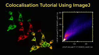
colocalisation tutorial using imagej
-
 7:18
7:18
fiji (imagej): quantification of 3d images - measuring number, volume, intensity, etc. on z stacks
-
 40:56
40:56
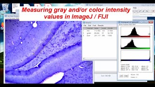
measuring gray & color intensity with imagej / fiji (see description)
-
 47:12
47:12
comparing color intensities between brightfield images in imagej / fiji (2023)
-
 5:40
5:40
how to count the number of seeds with different colors using imagej software
-
 1:01
1:01
fiji for quantification: area of stain