z stack image of hela cells labeled with celllightstm reagents
Published 10 years ago • 2K plays • Length 0:06Download video MP4
Download video MP3
Similar videos
-
 1:06
1:06
time lapse imaging of hela cells
-
 0:37
0:37
time lapse imaging of dividing hela cells
-
 2:57
2:57
create z-stacks using the evos fl auto 2 microscope
-
 0:24
0:24
time lapse imaging of hela cells treated with latrunculin a
-
 0:05
0:05
time-lapse with qtracker® 655
-
 0:32
0:32
3d-animation of hela cells mounted in prolong glass using structured illumination microscopy
-
 0:10
0:10
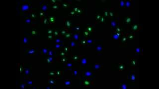
viability determination of hela cells using readyprobes® cell viability imaging kit (blue/green)
-
 8:10
8:10
your textbooks are wrong, this is what cells actually look like
-
 0:56
0:56
cell division under microscope
-
 47:40
47:40
best practices: 5 steps to live-cell imaging
-
 3:01
3:01
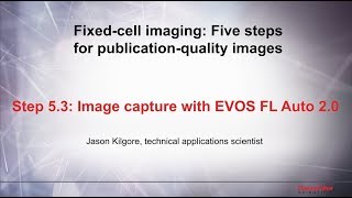
5.3 image capture with evos fl auto 2.0–fixed cell imaging: 5 steps for publication-quality images
-
 0:07
0:07
3-d rotation of histone and microtubules in a mitotic hela cell
-
 2:15
2:15
reviewing cell images on the evos fl auto 2 microscope
-
 0:16
0:16
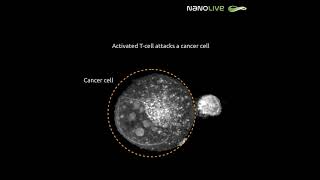
activated t cell attacks a cancer cell
-
 0:13
0:13
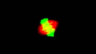
time lapse imaging of actin and talin
-
 0:17
0:17
live cell imaging with evos® fl auto imaging system
-
 3:42
3:42
fluorescence microscope demo video – evos fl
-
 0:31
0:31
hela cells - high frame timelapse, label free 3d live cell imaging
-
 0:14
0:14
time lapse of co-culture of hela cells together with 3t3 cells - cytosmart lux3 fl
-
 0:08
0:08
time lapse imaging of bpaec cells treated with menadione
-
 0:51
0:51
live dead cell imaging kit on the evos auto imaging system