how to measure cell size and shade a sub population of cells with a microscope image in imagej
Published 2 weeks ago • 52 plays • Length 7:20Download video MP4
Download video MP3
Similar videos
-
 9:42
9:42
how to measure fluorescence intensity within multiple regions of an image in imagej
-
 10:43
10:43
how to count objects in image using imagj| counting cells in imagej| imagej cell counter
-
 3:43
3:43
measuring images with imagej
-
 6:56
6:56
how to use imagej software to count cell numbers, analyze area and the intensity
-
 6:11
6:11
how to count cells using imagej | how to count cells in imagej | imagej cell counting |
-
 41:36
41:36
a beginners guide to imagej (and fiji)
-
 4:59
4:59
fiji (imagej): quantification of 2d images (measuring area, intensity, etc.)
-
 5:16
5:16
fiji for quantification: cell segmentation
-
 4:36
4:36
using imagej to measure the mean staining intensity of two different images
-
 8:24
8:24
part 6. data (image) analysis: image j to determine area of lipid droplets
-
 16:28
16:28
how electron microscope works
-
 9:05
9:05
how to set scale bar in imagej on microscope and camera images | image calibration
-
 8:36
8:36
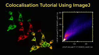
colocalisation tutorial using imagej
-
 9:08
9:08
batch processing in imagej, automatic image processing with a single click|microscope image analysis
-
 9:06
9:06
how to plot a line graph in imagej or fiji | intensity profile plot for microscope image
-
 9:34
9:34
using imagej to measure cell number and cross-sectional area of confocal images
-
![fiji (imagej): counting foci or puncta [difference of gaussians]](https://i.ytimg.com/vi/5z-BhcpiXp0/mqdefault.jpg) 5:20
5:20
fiji (imagej): counting foci or puncta [difference of gaussians]
-
 5:16
5:16
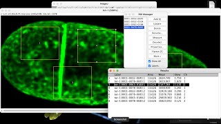
fiji (imagej): using 3d objects counter and 3d manager to measure cells in a z stack
-
 21:08
21:08
image j calibration with a stage micrometer
-
 11:01
11:01
how to quantify diseased leaf area in imagej| measure lesion size on leaf | imagej area measurement
-
 3:37
3:37
separating colors in light microscopy images: color deconvolution in imagej/fiji
-
 5:30
5:30
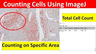
cell counting using imagej